Chapter 9: Control and Co-ordination
Multiple choice question.
HSC Biology Maharashtra State Board
HSC Biology Board Paper With Solutions
- JULY 2025
- MARCH 2025
- MARCH 2025 (Marathi)
- MARCH 2025 (Hindi)
- JULY 2024
- MARCH 2024
- MARCH 2024 (Hindi)
- JULY 2023
- MARCH 2023
- JULY 2022
- MARCH 2022
- MARCH 2020
- JULY 2019
- MARCH 2019
- JULY 2017
- MARCH 2017
- JULY 2016
- MARCH 2016
- JULY 2015
- MARCH 2015
- OCTOBER 2014
- MARCH 2014
- OCTOBER 2013
- MARCH 2013
Chapterwise List - Complete Bookback Solution.
- Chapter 1: Reproduction in Lower and Higher Plants
- Chapter 2: Reproduction in Lower and Higher Animals
- Chapter 3: Inheritance and Variation
- Chapter 4: Molecular Basis of Inheritance
- Chapter 5: Origin and Evolution of Life
- Chapter 6: Plant Water Relation
- Chapter 7: Plant Growth and Mineral Nutrition
- Chapter 8: Respiration and Circulation
- Chapter 9: Control and Coordination
- Chapter 10: Human Health and Diseases
- Chapter 11: Enhancement of Food Production
- Chapter 12: Biotechnology
- Chapter 13: Organisms and Populations
- Chapter 14: Ecosystems and Energy Flow
- Chapter 15: Biodiversity, Conservation and Environmental Issues
Very very short answer question.
Near the centre of the midbrain is a mass of grey matter scattered within the white matter. It is called the red nucleus. It plays an important role in controlling posture and muscle tone, modifying some motor activities, and motor coordination.
The corpora quadrigemina are four rounded elevations on the dorsal surface of the midbrain. The two superior colliculi are involved in visual reflexes and the two inferior colliculi are relay centres for auditory reflexes that operate when it is necessary to move the head to hear sounds more distinctly.
Cerebellum: It is the second-largest part of the brain and consists of two lateral hemispheres and a central vermis. It is composed of white matter with a thin layer of grey matter, the cortex. The white matter intermixes with the grey matter and shows a tree-like pattern called arborvitae. The surface of the cerebellum shows convolutions (gyri and sulci) a number of nuclei lie deep within each lateral or cerebellar hemisphere. Over 30 million neurons lie in the cortex. Three pairs of myelinated nerve bundles called cerebellar peduncles to connect the cerebellum to the other parts of the CNS.
Functions: It is an important centre that maintains the equilibrium of body, posture, balancing orientation, moderation of voluntary movements, maintenance of muscle tone. It is a regulatory center for neuromuscular activities and controls rapid activities like walking, running, speaking, etc. All activities of the cerebellum are involuntary (though may involve learning in early stages).
Middle ear: It consists of a chain of three ear ossicles called Malleus (hammer), Incus (anvil), and Stapes (stirrup-the smallest bone). On receiving the vibrations from the tympanic membrane, the ear ossicles amplify the vibrations and transfer these to the cochlea.
A short Eustachian tube connects the middle ear to the pharynx. It equalizes air pressure on both sides of the eardrum.
Progesterone
Placenta
Steroids
Excessive secretion of Growth Hormone causes abnormal elongation of long bones of arms and legs and of the lower jaw.
Melatonin
Thymus gland
| 1. Neurons | a. Earthworm |
| 2. Ladder-type | b. Hydra |
| 3. Ganglion | c. Flatworm |
| 4. Nerve net | d. Human |
| 1. Neurons | d. Human |
| 2. Ladder-type | c. Flatworm |
| 3. Ganglion | a. Earthworm |
| 4. Nerve net | b. Hydra |
Islets of Langerhans are endocrine cells of the pancreas. They are four types of cells in Islets of Langerhans which have an endocrine role i.e. they secrete hormones.
i. Alpha cells (α cells): They constitute 20% of Islets of Langerhans. They secrete hormone glucagon. Glucagon stimulates glycogenolysis (the breakdown of glycogen) in the liver which causes hyperglycemia.
ii. Beta cells (β cells): They constitute 70% of Islets of Langerhans. They secrete insulin which stimulates glycogenesis (formation of glycogen) in the liver and muscles. Insulin causes hypoglycemia by increasing the uptake of glucose by cells.
iii. Delta cells (δ cells): They constitute 10% of Islets of Langerhans. These cells secrete somatostatin which inhibits the secretion of insulin and glucagon. It also lowers gastric secretions, motility, and absorption in the digestive tract. Somatostatin inhibits the release of growth hormone.
iv. PP cells or F cells: These cells secrete pancreatic polypeptide (PP) and inhibit the release of pancreatic juice.
The group of hormones secreted by testis is androgens such as testosterone.
Functions of androgens:
i. It is also responsible for the appearance of secondary sexual characters such as facial and pubic hair, deepening of the voice, broadening of shoulders, male aggressiveness, etc.
ii. It involves the development of testis.
This condition causes excessive micturition or polyuria, polydipsia (increased thirst), etc.
Short answer question
i. When Rakesh fell down from his motorbike the inner membranes called meninges protected his brain from injury.
ii. These meninges form a protective covering around the brain and spinal cord. They act as shock absorbers.
i. The medulla oblongata is a part of the brain stem.
ii. It controls involuntary vital functions like heartbeat, respiration, vasomotor activities, and peristalsis.
iii. It also controls non-vital reflex activities like coughing, sneezing, swallowing, vomiting, yawning, etc.
iv. Thus, damage or injury to medulla oblongata may disrupt these vital functions. Therefore, injury to medulla oblongata may prove fatal.
i. Heart: Sympathetic nervous system accelerates the heartbeat whereas the parasympathetic nervous system decelerates the heartbeat.
ii. Urinary bladder: Sympathetic nervous system inhibits bladder contraction whereas the parasympathetic nervous system stimulates bladder contraction.
i. Mr. Kothari may be suffering from Graves’ disease.
ii. It is caused due to increased levels of thyroid hormone or hyperthyroidism.
i. Excitability/Irritability: Nerve fibres have polarized membranes, thus they have the ability to perceive stimulus and enter into a state of activity.
ii. Conductivity: It is the ability of the nerve to transmit impulses along the whole length of the axon.
iii. Stimulus: It is any detectable, physical, chemical, electrical change in the external or internal environment which brings about excitation in a nerve/muscle/organ/organism. A stimulus must have a minimum intensity called threshold stimulus, in order to be effective. The subliminal (weak) stimulus will have no effect while the supraliminal (strong) stimulus will produce the same degree of impulse as the threshold stimulus.
iv. Summation effect: A single subliminal stimulus will have no effect but when many such weak stimuli are given again and again they may produce an impulse due to summation of effects.
v. All or none law: The nerve will either conduct the impulse along its entire length or will not conduct the impulse at all. This occurs in the case of a subliminal or weak stimulus.
vi. Refractory period: It is the time interval (about a millisecond) during which a nerve fails to respond to a second stimulus even if it is strong.
vii. Synaptic delay: The impulse takes about 0.3 to 0.5 milliseconds to cross a synapse. It is required for the release of neurotransmitters from the axon terminal and excitation in the dendron of the next neuron.
viii. Synaptic fatigue: The transmission of nerve impulses across the synapse stops temporarily due to the depletion of the neurotransmitter.
ix. Velocity: The rate of transmission of impulse is higher in long and thick nerves. It is higher in homeotherms than in poikilotherms. The velocity of transmission is higher in voluntary fibres (100 - 120 m/s in man) as compared to autonomic or involuntary nerves (10-20 m/s). In medullated nerve fibre, the velocity of transmission is higher as an impulse has to jump from one node of Ranvier to the next.
The tongue detects the sensation of taste due to gustatoreceptors.
i. Site of production: Secretin, gastrin, and cholecystokinin are secreted in the gastrointestinal tract
ii. Functions:
Secretin: It is responsible for the secretion of pancreatic juice from the pancreas and bile from the liver.
Gastrin: It stimulates gastric glands to produce gastric juice.
Cholecystokinin CCK/ Pancreozymin PZ: It stimulates the pancreas to release enzymes and also stimulates the gallbladder to release bile
i. The patient may be suffering from myxoedema.
ii. It is caused due to the deficiency of thyroid hormones (hypothyroidism) in adults.
iii. Care: Patients should take prescribed medications regularly and eat food rich in iodine.
The pituitary gland located just below the hypothalamus.
Hormones secreted by anterior pituitary:
i. Somatotropic Hormone (STH) / Somatotropin / Growth Hormone (GH):
The secretion of GH is high till puberty later its secretion becomes low. However, it is continuously secreted throughout life for repair and replacement of body tissue or cells.
Functions:
a. It stimulates the growth of the body and the development of all tissues.
b. It accelerates protein synthesis and cell division.
c. It stimulates the release of growth hormone.
ii. Thyroid Stimulating Hormone (TSH) / Thyrotropin:
Function:
It stimulates the thyroid gland to produce the hormone thyroxine.
iii. Adrenocorticotropic Hormone (ACTH) / Adrenocorticotropin:
Functions:
a. It stimulates the adrenal cortex to produce its hormones.
b. It maintains the functioning of the adrenal cortex.
iv. Prolactin / Luteotropin/ Mammotropin :
The secretion of this hormone is regulated by PIF (Prolactin inhibiting factor) of the hypothalamus.
Functions:
a. Activates the growth of mammary glands during pregnancy (mammotropin).
b. Stimulates milk production and secretion of milk (lactogenic) by the mammary gland after childbirth.
v. Gonadotropins:
a. Follicle Stimulating Hormone (FSH): In males, it stimulates the development of seminiferous tubules. In females, it stimulates the growth of ovarian follicles.
b. Luteinizing hormone (LH): LH induces the ruptured follicles to develop into corpus luteum and for the production of progesterone FSH and LH are responsible for the stimulation of ovaries to produce estrogen.
c. ICSH: In males, it stimulates the testes to produce the androgen called testosterone. Testosterone is responsible for the development of secondary sexual characters.
Islets of Langerhans are endocrine cells of the pancreas.
They are four types of cells in Islets of Langerhans which have an endocrine role i.e. they secrete hormones.
i. Alpha cells (α cells): They constitute 20% of Islets of Langerhans. They secrete hormone glucagon. Glucagon stimulates glycogenolysis (the breakdown of glycogen) in the liver which causes hyperglycemia.
ii. Beta cells (β cells): They constitute 70% of Islets of Langerhans. They secrete insulin which stimulates glycogenesis (formation of glycogen) in the liver and muscles. Insulin causes hypoglycemia by increasing the uptake of glucose by cells.
iii. Delta cells (δ cells): They constitute 10% of Islets of Langerhans. These cells secrete somatostatin which inhibits the secretion of insulin and glucagon. It also lowers gastric secretions, motility, and absorption in the digestive tract. Somatostatin inhibits the release of growth hormone.
iv. PP cells or F cells: These cells secrete pancreatic polypeptide (PP) and inhibit the release of pancreatic juice.
Hypersecretion of thyroid hormones: It is caused by an increase in the levels of thyroid hormones. This increases metabolic rate, sensitivity, sweating, flushing, rapid respiration, bulging of eyeballs, and affects various physiological activities. Graves’ disease: Hyperthyroidism in adults results in this disorder. It is characterised by protruding eyeballs, increased BMR, and weight loss. Increased BMR produces a range of effects like increased heartbeat, increased B.P., higher body temperature, nervousness, irritability, and tremor of fingers.
Simple goitre: It is iodine deficiency goitre. Iodine is required for the synthesis of thyroid hormone and if there is a deficiency of iodine in the diet, it causes enlargement of the thyroid gland leading to simple goitre. This disease is common in hilly areas. Addition of iodine to table salt prevents this disease. The size of the thyroid gland is increased but the total output of thyroxine is decreased.
Ovaries secrete the following hormones:
i. Progesterone: It is secreted by the corpus luteum of the ovary after ovulation. It is essential for the thickening of the uterine endometrium, thus preparing the uterus for implantation of the fertilized ovum. It is responsible for the development of mammary glands during pregnancy. It inhibits uterine contractions during pregnancy.
ii. Oestrogen: It is secreted by developing follicles. Estradiol is the main oestrogen. It is responsible for the development of secondary sexual characters in females.
iii. Relaxin: It is secreted by the corpus luteum of the ovary at the end of the gestation period. It relaxes the cervix of the pregnant female and the ligaments of the pelvic girdle to ease out the birth process.
iv. Inhibin: It is secreted by the corpus luteum. Inhibin inhibits FSH and GnRH production.
Answer the following.
| Location | Cell Type | Function |
|---|---|---|
| PNS | ___________ | Produce myelin sheath |
| PNS | Satellite cells | _____________ |
| ___________ | Oligodendrocytes | Form myelin sheath around central axon |
| CNS | ___________ | Phagocytose pathogens |
| CNS | ___________ | Form the epithelial lining of brain cavities and central canal. |
| Location | Cell Type | Function |
|---|---|---|
| PNS | Schwann cells | Produce myelin sheath |
| PNS | Satellite cells | Support the function of neurons |
| CNS | Oligodendrocytes | Form myelin sheath around the central axon |
| CNS | Microglia or brain macrophages | Phagocytose pathogens |
| CNS | Ependymal cells | Form the epithelial lining of brain cavities and central canal. |
Long answer question.
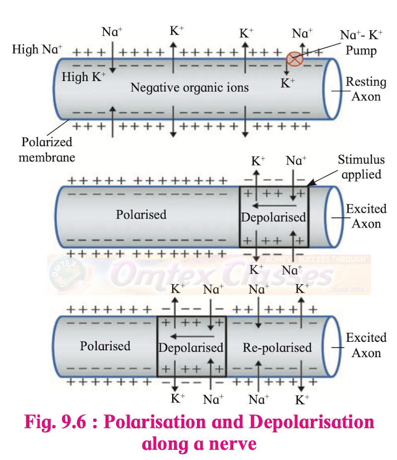
Polarisation and Depolarisation along a nerve
i. The nerve impulse is a wave of bioelectrical or electrochemical disturbances passing along a neuron.
ii. Neurons have a charged cellular membrane with a voltage that is different on the outer and inner side of the membrane. The plasma membrane separates the outer and inner solutions of different chemical compounds but having approximately the same total number of ions.
iii. The external tissue fluid has both Na+ and K+ but there is a predominance of Na+ and Cl-, while K+ is predominant within the fibre or in the intracellular fluid. This condition of a resting nerve is also called a polarised state.
iv. The polarized state of a neuron is established by maintaining an excess of Na+ on the outer side. On the inside, there is an excess of K+ along with large negatively charged protein molecules and nucleic acid.
v. Some amount of Na+ and K+ always leaks across the membrane. The Na+/K+ pump actively maintains the ionic gradient across the resting membrane. The sodium pump or Na-K allows the entry of K+ inside the membrane and exit of Na+.
vi. The difference in the distribution of Na+ and K+ on the two sides of the membrane produces a potential difference of – 50 to –100 millivolts (average is – 70 millivolts).
vii. The potential difference seen in a resting nerve is thus called resting potential (–70 millivolts) and it is mainly due to differential permeability of the resting membrane, which is much more permeable to K+ than to Na+. This results in slightly more K+ diffusing out than Na+ moving inside and causing a slight difference in polarity.
viii. Also, ions like negatively charged proteins and nucleic acids inside the cell make the overall charge negative on the inside and positive charge on the outside. The nerve membrane not only has leakage channels but also has many gated channels for Na+ /K+. These are also called voltage-gated channels. These channels enable the neuron to change its membrane potential to active potential in response to stimuli. The Na+ /K+ gated channels are separate so the transport of both these ions is separately done. However, during resting potential, both these gates are closed and the membrane resting potential is maintained.
ix. The resting potential of the membrane is maintained unless the stimulus reaches the neuron. Any change or disturbance to the membrane will cause Na+ to enter into the membrane and lower the potential difference (lesser than –70 millivolts). Thus, the membrane becomes more permeable to Na+
x. During resting potential, both gates are closed, and resting potential is maintained. However, during depolarization, the Na+ gates open and the K+ gates remain closed. This causes Na+ to rush into the axon and bring about depolarisation (opposite of polarity).
xi. The Extra Cellular Fluid (ECF) becomes electronegative with respect to the inner membrane which becomes electropositive. The value of action potential reaches +30 millivolts to +60 millivolts. This triggers depolarization in the next part while it itself starts going to repolarisation.

Structure of the human ear

A sectional view of the human eye
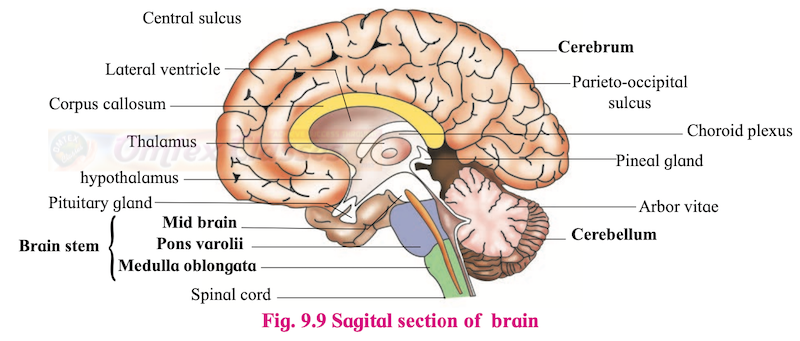
Sagital section of the brain

Multipolar Neuron
Long Answer Questions
Answer the question after observing the diagram given below.

Synaptic vesicles contain neurotransmitter molecules.
A neurotransmitter is released by the process of exocytosis.
a. When a neuron receives an impulse, it passes it to the next neuron. The impulse travels along the axon of the pre-synaptic neuron to the axon terminal.
b. Pre-synaptic neurons or axons have synaptic knobs at their ends or terminals. These synaptic knobs have synaptic vesicles that contain neurotransmitter molecules.
c. When the impulse reaches a synaptic knob, Ca+ channels open and Ca+ ions diffuse inward from the extracellular fluid.
d. This causes the release of neurotransmitters that bind to the receptors of the post synaptic cell.
e. The neurotransmitter is destroyed by the enzyme cholinesterase. A new impulse/ next impulse is generated and conducted to the synaptic gap.
a. A pre synaptic neuron when receives an impulse, releases a neurotransmitter into the synaptic cleft which is required to cross the gap between the axon terminal and the next neuron.
b. For further transmission of impulse, a pre-synaptic neuron is required that initiates the release of neurotransmitters that facilitate the movement of impulses across synapses.
The ligand-gated ion channel is responsible for the transmission of the impulse.
i. The reflex pathway comprises at least one afferent neuron (receptor) and one efferent (effector or excitor) neuron appropriately arranged in a series.
ii. The afferent neuron receives a signal from a sensory organ and transmits the impulse via a dorsal nerve root into the CNS (at the level of the spinal cord).
iii. The efferent neuron then carries signals from CNS to the effector. The stimulus and response thus form a reflex arc as shown below in the knee jerk reflex.

The sympathetic nervous system controls body activities during fight, fright, or flight situations. It activates the release of the hormones adrenaline and nor-adrenaline due to which the heartbeat increases, hands and feet become cold.
Thyroid gland atrophy causes hypothyroidism.
For effects of thyroid gland atrophy on the human body:
Hyposecretion of thyroid hormone: It is caused by a deficiency of thyroid hormones or removal of the thyroid gland (Thyroidectomy).
a. Cretinism: It is caused due to deficiency of thyroid hormones in infants. A cretin (individual suffering from cretinism) has reduced BMR and low oxidation. They are short-statured because the skeleton fails to grow. They are mentally retarded, show stunted growth and delayed puberty. They show dry skin, thick tongue, prolonged neonatal jaundice, lethargy and constipation. This can be treated by early administration of thyroid hormones.
b. Myxoedema: It is caused due to the deficiency of thyroid hormones in adults. It is characterised by a peculiar thickening and puffiness of skin and subcutaneous tissue particularly of the face and extremities. The patient lacks alertness, intelligence. The patient suffers from slow heart rate, low B.P., low body temperature (feels cold) and stunted sexual development.
c. Simple goitre: It is iodine deficiency goitre. Iodine is required for the synthesis of thyroid hormone and if there is a deficiency of iodine in the diet, it causes enlargement of the thyroid gland leading to simple goitre. This disease is common in hilly areas. Addition of iodine to table salt prevents this disease. The size of the thyroid gland is increased but the total output of thyroxine is decreased.
a. Growth of thyroid and secretion of thyroxine.
b. Helps in relaxing pubic ligaments to facilitate the easy birth of young ones.
c. Stimulate intestinal glands to secrete intestinal juice.
d. Controls calcium level in the blood
e. Control tubular absorption of water in kidneys.
f. Urinary elimination of water.
g. Sodium and potassium ion metabolism.
h. Basal Metabolic rate.
i. Uterine contraction.
j. Heartbeat and blood pressure.
k. Secretion of growth hormone.
l. Maturation of Graafian follicle.
Solution:| Functions | Hormones | Glands |
|---|---|---|
| Growth of thyroid and secretion of thyroxine | TSH (Thyroid-stimulating hormone) | Pituitary |
| Helps in relaxing pubic ligaments to facilitate the easy birth of young ones. | Relaxin | Ovaries |
| Stimulate intestinal glands to secrete intestinal juice | Cholecystokinin | Gastrointestinal tract |
| Controls calcium level in the blood | Calcitonin, Parathormone | Thyroid, parathyroid respectively |
| Controls tubular absorption of water in kidneys | ADH/ Vasopressin | Hypothalamus |
| Urinary elimination of water | Antidiuretic hormone | Hypothalamus |
| Sodium and potassium ion metabolism | Aldosterone (mineralocorticoid) | Adrenal cortex |
| Basal Metabolic rate | Thyroxine | Thyroid |
| Uterine contraction | Oxytocin | Hypothalamus |
| Heartbeat and blood pressure | Adrenaline, nor adrenaline | Adrenal medulla |
| Secretion of growth hormone | Somatotropin | Pituitary |
| Maturation of Graafian follicle | LH (Luteinizing hormone) | Pituitary |
i. The hypothalamus controls the secretory activity of the pituitary gland (anterior pituitary) by producing, releasing, and inhibiting hormones.
ii. Anterior pituitary and intermediate lobes are connected to the hypothalamus through the hypophyseal portal system. Various hormones secreted by the hypothalamus reach the pituitary gland through the hypophyseal portal system.
iii. The portal vein collects blood from various parts of the hypothalamus and opens into the anterior lobe of the pituitary. From the pituitary, the vein finally carries the blood into the superior vena cava. It helps in the feedback mechanism for hormonal control.
iv. Also, a negative feedback mechanism takes place in the form of hormones released by the target glands to decrease the secretion of the pituitary gland.
v. In such a negative feedback mechanism, the secretion of ACTH, TSH, and gonadotropins (FSH and LH) decreases when their target gland hormone levels rise.
e.g. Adrenocorticotropic hormone (ACTH) stimulates the cortex of the adrenal gland to secrete glucocorticoids, mainly cortisol. In turn, an elevated blood level of cortisol decreases secretion of both corticotropin and corticotropin-releasing hormone (CRH) by suppressing the activity of the anterior pituitary corticotrophs and neurosecretory cells.
Adenohypophysis: It is an outgrowth from the roof of the buccal cavity. This outgrowth is called Rathke’s pouch. It grows upwards towards the brain. It is the larger lobe of the pituitary gland. It is a highly cellular and vascular part of the pituitary gland. It contains various types of epitheloid secretory cells, acidophils, basophils, chromatophores. It is further divided into three parts - Pars distalis, pars tuberalis, and pars intermedia. Pars intermedia is poorly developed in human beings. It is a small reduced part lying in the cleft between the anterior and posterior lobe. It secretes Melanocyte Stimulating Hormone (MSH) in some lower vertebrates. MSH stimulates the dispersion of melanin granules in melanocytes and is responsible for skin pigmentation.
The pituitary gland located just below the hypothalamus.
Hormones secreted by anterior pituitary:
i. Somatotropic Hormone (STH) / Somatotropin / Growth Hormone (GH): The secretion of GH is high till puberty later its secretion becomes low. However, it is continuously secreted throughout life for repair and replacement of body tissue or cells.
Functions:
a. It stimulates the growth of the body and the development of all tissues.
b. It accelerates protein synthesis and cell division.
c. It stimulates the release of growth hormone.
ii. Thyroid Stimulating Hormone (TSH) / Thyrotropin:
Function:
It stimulates the thyroid gland to produce the hormone thyroxine.
iii. Adrenocorticotropic Hormone (ACTH) / Adrenocorticotropin:
Functions:
a. It stimulates the adrenal cortex to produce its hormones.
b. It maintains the functioning of the adrenal cortex.
iv. Prolactin / Luteotropin/ Mammotropin: The secretion of this hormone is regulated by PIF (Prolactin inhibiting factor) of the hypothalamus.
Functions:
a. Activates the growth of mammary glands during pregnancy (mammotropin).
b. Stimulates milk production and secretion of milk (lactogenic) by the mammary gland after childbirth.
v. Gonadotropins:
a. Follicle Stimulating Hormone (FSH): In males, it stimulates the development of seminiferous tubules. In females, it stimulates the growth of ovarian follicles.
b. Luteinizing hormone (LH): LH induces the ruptured follicles to develop into corpus luteum and for the production of progesterone FSH and LH are responsible for the stimulation of ovaries to produce estrogen.
c. ICSH: In males, it stimulates the testes to produce the androgen called testosterone. Testosterone is responsible for the development of secondary sexual characters.
Disorders of the adrenal gland:
i. Addison’s disease: It is caused due to hypersecretion of glucocorticoids (hormone secreted adrenal cortex). It is characterized by low blood sugar, low Na+, and high K+ concentration in plasma increased loss of Na+ and water in urine. It leads to weight loss, weakness, nausea, vomiting, and diarrhoea.
ii. Cushing’s disease: It is caused due to Hyposecretion of mineralocorticoids (a hormone secreted by the adrenal cortex) It leads to high blood sugar levels, excretion of glucose in the urine, rise in Na+ level in the blood, high blood pressure, obesity, and wasting of limb muscles.
Hormones are released in very minute quantities. They produce their effect on the target cells by binding to hormone receptors. The hormone receptors are present on the cell membrane (i.e. membrane receptors) or maybe intracellular receptors.
i. Mode of hormone action through membrane receptors:
a. Hormones like catecholamines, peptide, and polypeptide hormones are not lipid-soluble and they cannot enter their target cells through the plasma membrane. These non-steroid water-soluble hormones interact with surface receptors and initiate metabolic activity.
b. Molecules of amino acid derivatives, peptide hormones bind to specific receptor molecules located on the plasma membrane.
c. The hormone-receptor complex causes the release of an enzyme adenylate cyclase from the receptor site. This enzyme forms cyclic AMP from ATP of the cell.
d. cAMP activates enzymatic actions. The hormone acts as the first messenger and the cAMP acts as the second messenger.
e. Some other secondary messengers are Ca++, cGMP, and IP3 (Inositol triphosphate), etc.
ii. Mode of action through intracellular receptors:
a. Steroid and thyroid hormones are lipid-soluble and can easily pass through the plasma membrane of the target cell into the cytoplasm.
b. In the cytoplasm, they bind to specific intracellular receptors proteins forming a hormone-receptor complex that enters the nucleus.
c. The hormone-receptor complex binds to a specific regulatory site of DNA, in the nucleus.
d. The activated genes transcribe mRNA which directs protein synthesis and enzymes in the cytoplasm.
e. The action of lipid-soluble hormones is slow but long-lasting,

Mechanism of hormone action through membrane receptor
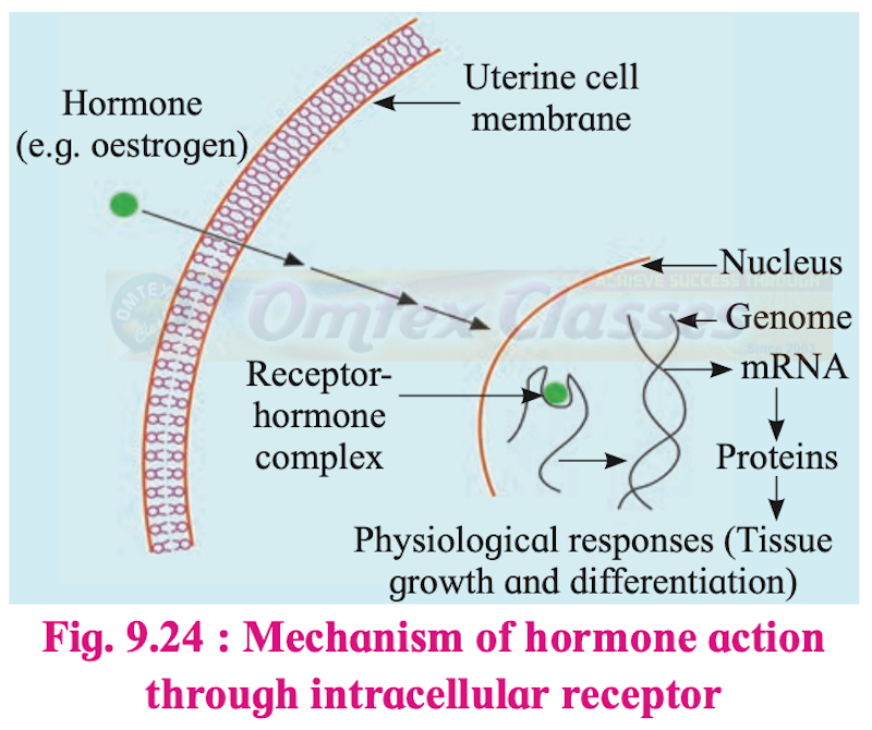
Mechanism of hormone action through intracellular receptor
Disorders of the thyroid gland are caused due to hypersecretion and hyposecretion of thyroid hormones.
i. Hypersecretion of thyroid hormones: It is caused by an increase in the levels of thyroid hormones. This increases metabolic rate, sensitivity, sweating, flushing, rapid respiration, bulging of eyeballs, and affects various physiological activities.
Graves’ disease: Hyperthyroidism in adults results in this disorder. It is characterised by protruding eyeballs, increased BMR, and weight loss. Increased BMR produces a range of effects like increased heartbeat, increased B.P., higher body temperature, nervousness, irritability, and tremor of fingers.
ii. Hyposecretion of thyroid hormone: It is caused by a deficiency of thyroid hormones or removal of the thyroid gland (Thyroidectomy).
a. Cretinism: It is caused due to deficiency of thyroid hormones in infants. A cretin (individual suffering from cretinism) has reduced BMR and low oxidation. They are short-statured because the skeleton fails to grow. They are mentally retarded, show stunted growth and delayed puberty. They show dry skin, thick tongue, prolonged neonatal jaundice, lethargy and constipation. This can be treated by early administration of thyroid hormones.
b. Myxoedema: It is caused due to the deficiency of thyroid hormones in adults. It is characterised by a peculiar thickening and puffiness of skin and subcutaneous tissue particularly of the face and extremities. The patient lacks alertness, intelligence. The patient suffers from slow heart rate, low B.P., low body temperature (feels cold) and stunted sexual development.
c. Simple goitre: It is iodine deficiency goitre. Iodine is required for the synthesis of thyroid hormone and if there is a deficiency of iodine in the diet, it causes enlargement of the thyroid gland leading to simple goitre. This disease is common in hilly areas. Addition of iodine to table salt prevents this disease. The size of the thyroid gland is increased but the total output of thyroxine is decreased.
iii. Hyposecretion of thyroid hormones in pregnant females causes defective development and maturation of growing baby.